
Biology
Given below is the structure of a human eye. Read the information below the diagram and fill in the blanks:
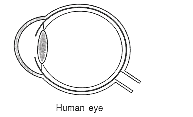
Eyes are the sense organs located in deep sockets on the front side of the head. Eyes can receive the light stimulus and hence considered as photoreceptors. Eyes are protected with eyelids, eyebrows and tear glands, etc. Eyes are visionary in function.
The wall of the eyeball is composed of three concentric layers. The middle layer of the eyeball is (a) ……………, richly supplied with blood vessels whereas the innermost layer is (b) …………… which contains two types of sensory cells. (c) …………… is the extension of the middle layer of the eyeball which regulates the size of the (d) …………… . The image formed on the yellow spot of the eye is the (e) ……………
Sense Organs
18 Likes
Answer
(a) Choroid
(b) Retina
(c) Iris
(d) Pupil
(e) brightest
Answered By
15 Likes
Related Questions
Note the relationship between the first two words and suggest the suitable word/words for the fourth place.
(a) Cones : Iodopsin :: Rods: …………… .
(b) Eyes : Photoreceptors :: Ears : …………… .
(c) Ears : Auditory nerve :: Eyes : …………… .
(d) Ear pinna : Auricle :: Inner ear : …………… .
(e) Semi-circular canal : Ampulla :: Cochlea : …………… .
Match the terms in column I with those in column II and write down the matching pairs.
Column I Column II Conjunctiva Viral infection Cornea Ciliary body Choroid Spiral-shaped Cochlea Transparent epithelium Conjunctivitis Suspensory ligament Contains melanin Transparent but appears black State whether the following statements are true (T) or false (F). If false, correct them by changing any one single word in each.
(a) Deafness is caused due to rupturing of the pinna.
(b) Semicircular canals are concerned with static (positional) balance.
Where are the following located?
(a) Yellow spot
(b) Lacrimal gland
(c) Organ of Corti
(d) Eustachian canal
(e) Incus